

New confocal microscope offers more hands-on learning and research opportunities
Staying updated on the latest research and having access to vital resources is essential to any successful career. This is especially true in scientific careers such as neuroscience, where technology and novel ideas rapidly and regularly change. Middlebury College understands this. That’s why several of its faculty members recently worked together to provide their students with a crucial piece of scientific equipment: a confocal microscope.
The endeavor was proposed by five Middlebury faculty members: Clinton Cave, a neuroscience professor; Amanda Crocker, a neuroscience professor; Catherine Combelles, a biology professor; Michael Durst, a physics professor; and Mark Spritzer, a biology professor. The microscope was purchased via a grant awarded by the National Science Foundation (NSF) in 2020. The NSF was established in 1950 by Congress to promote the progress of science, advance national health, and secure national defense.
“Within our microscopy facility, there are 4 compound microscopes with epi-fluorescent capabilities, and several faculty members have good research microscopes in their labs. We also have one nice scanning electron microscope,” said Spritzer. “But none of this equipment has the same abilities as the confocal microscope.”
A confocal microscope can focus light through a narrow pinhole. The pinhole centralizes the light, narrowing the image plane. This eliminates background blurring and illuminates a sample, specimen, or object, offering a detailed image. A conventional fluorescent microscope does not possess a pinhole. Thus, it evenly distributes light across a wide plane which results in a lower-resolution image.
Confocal microscopes are used to better understand relationships between the structure and function of cells within a tissue, such as the relationship between neurons. It also offers 3-D images of cells and intracellular structures. This insight is used to support basic life science research, drug development, and medical research.
Prior to being awarded their grant, Middlebury’s faculty and students traveled to use a confocal microscope. The closest one was at the University of Vermont in Burlington—about an hour’s drive away from Middlebury. Middlebury had to pay to use it. For example, prior to this grant, the University of Vermont (UVM) owned the only confocal microscope in the entire state of Vermont. Yet, the microscope costs about $55/hour to use. Additionally, because students require training and supervision, it costs more if Middlebury students needed to use UVM’s equipment (~$50-$100/hr.)
In addition, according to the team’s grant proposal, 13 former neuroscience students, who did either summer and/or senior research at Middlebury, expressed that experience with a confocal during their undergraduate careers would have helped their resume. Ten of these students felt it would have helped them acquire a research technician position or aided in their acceptance to graduate school.
This feedback, alongside travel expenses, logistics, and scheduling difficulties, encouraged the team to apply for an NSF grant. The grant was used to purchase and put a microscope directly on Middlebury’s campus, which helped to overall reduce costs and offered more opportunities to do research with state-of-the-art equipment. Middlebury applied twice for an NSF grant—once in 2017 and once in 2018. However, it was not awarded the grant until 2020.
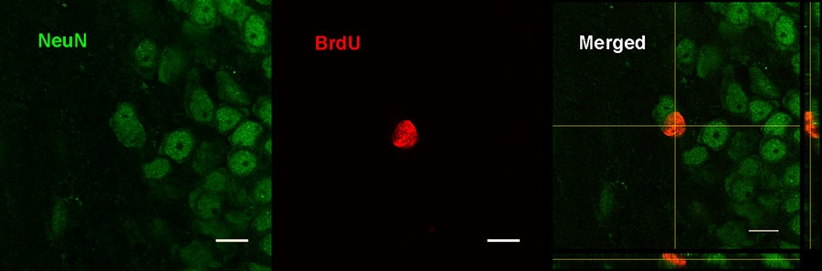
“The main change that we made in the [2020] submission was to include better quality images, which better demonstrated the excellent images that we could obtain with the Olympus microscope,” explained Spritzer. “Another critical component was being able to demonstrate that Middlebury College provides opportunities for groups of students (race and socioeconomic status) who are under-represented in the sciences.”
Within our microscopy facility, there are 4 compound microscopes with epi-fluorescent capabilities, and several faculty members have good research microscopes in their labs. We also have one nice scanning electron microscope,” said Spritzer. “But none of this equipment has the same abilities as the confocal microscope.
According to the Middlebury’s Assessment and Institutional Research website, about 20% of Middlebury’s undergraduate class was awarded a natural science degree in 2019. This percentage rose to about 23% in 2020 and 2021. This makes natural science the second most awarded degree at Middlebury’s undergraduate program; social sciences degrees are number one.
Among these natural science degrees, certain majors have seen an upsurge. For instance, in 2019, 1.2% of the awarded degrees were Physics majors, 3.0% were Biology majors, and 3.1% were Molecular Biology and Biochemistry majors. By 2021, though, 2.5% majored in Physics, 3.4% in Biology, and 3.3% in Molecular Biology and Biochemistry. Among these students, 20-30% enter graduate school or health professional schools immediately after graduation.
To determine which microscope fit its needs, the faculty received presentations from three vendors (Olympus, Nikon, and Lecia). Then, Middlebury compared the advantages and disadvantages of each microscope’s features, price, and size to each other as well as the equipment at neighboring institutions. It also shared future projects that the microscope would support and garnered written support from nearby universities and colleges, including Norwich University and Castleton University, which now has access to Middlebury’s new microscope,
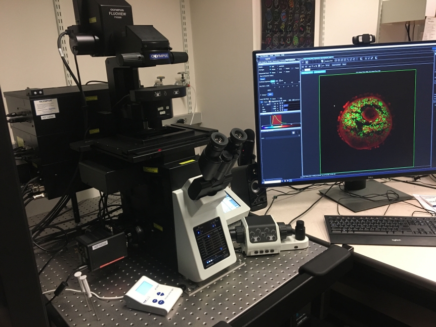
Eventually, it was decided that the Olympus Fluoview FV3000 was the best fit. The microscope is now located at McCardell Bicentennial Hall. McCardell Bicentennial Hall has been Middlebury’s multidisciplinary hub for scientific research for more than 20 years. It is home to six academic departments: Biology, Chemistry and Biochemistry, Geography, Earth and Climate Sciences, Physics, and Psychology. It’s the center for three major programs as well, including Environmental Studies, Neuroscience, and Molecular Biology and Biochemistry.
This instrumentation is truly elevating our science here at the college. Its versatility allows us to answer many kinds of research questions,” said Cave. “Last year, we were imaging the spatial distribution of reactive glial cells during neurodegeneration. Our most recent paper used the confocal microscope to detect single RNA molecules within individual cells. This summer we’ll be making timelapse movies of entire zebrafish embryos as they grow nervous systems.
Many of these listed majors are directly impacted by the available resources used at McCardell Bicentennial Hall. This includes the confocal microscope, which is now available to students working on independent study or thesis projects with professors.
For instance, The Crocker lab uses the microscope to identify the cellular changes in the brain after exposure to painful stimuli. The Durst lab uses it for photothermal imaging of absorbing structures in thick tissue. Cave’s lab uses the microscope to identify the subcellular localization of proteins within the developing spinal cord. Meanwhile, the Combelles lab uses it to study the specific areas or targets in ovarian follicles that are affected by exposure to endocrine-disrupting chemicals. Some images acquired from this microscope are already being published, too.
“One line of research in my laboratory is investigating how different hormones influence neuron growth in the adult brain. The goal is to develop drugs to help treat neurodegenerative diseases,” Spritzer said. “Collecting and analyzing brain tissue with a confocal microscope is essential for such projects. Over the years, my research lab had developed a large backlog of tissue samples that needed to be analyzed. Now, we are rapidly working through that tissue, which will likely lead to at least three scientific publications.”
“This instrumentation is truly elevating our science here at the college. Its versatility allows us to answer many kinds of research questions,” said Cave. “Last year, we were imaging the spatial distribution of reactive glial cells during neurodegeneration. Our most recent paper used the confocal microscope to detect single RNA molecules within individual cells. This summer we’ll be making timelapse movies of entire zebrafish embryos as they grow nervous systems.”
The list of courses impacted by this microscope is growing as well. Originally, the proposal team anticipated the microscope to be incorporated in at least seven Middlebury courses. However, that number has increased as the microscope offers more versatility and hands-on research opportunities. Some of the impacted courses include physics courses and three upper-level biology/neuroscience courses like Animal Physiology, Molecular Neurogenetics, and Methods in System Neuroscience.
This broader impact means that upwards of 120 students have direct hands-on access to this new microscope every year. Hence, this recent NSF grant has not only resulted in cost savings and eliminated logistical conundrums. It has also allowed Middlebury College to continue offering immersive learning experiences that prepare students for their future careers as next-generation researchers, who can magnify and shed light on our understanding of the world.